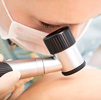
Integumentary System: All About Our Skin

This lesson contains affiliate links to products I have used and personally recommend. At no cost to you, I make a commission for purchases made through the links or advertisements. These commissions help to pay for the costs of the site and enable it to remain free for anyone who wants to use it.
Objectives:
-
The students will be able to explain the functions of the skin.
-
The students will be able to describe the three layers of skin.
-
The students will be able to describe some pathologies of the integumentary system.
-
The students will be able to explain the importance of keeping the skin clean.
Questions that encompasses the objective:
-
Think about your skin. Why do you think we have skin?
Prepare the Learner: Activating Prior Knowledge.
How will students prior knowledge be activated?
Warm up by asking students:
-
What do you know about your skin?
Common Core State Standards:
-
CCSS.ELA-LITERACY.SL.2.1
-
CCSS.ELA-LITERACY.SL.2.1 B
-
CCSS.ELA-LITERACY.SL.2.4
-
CCSS.ELA-LITERACY.W.2.2
-
CCSS.ELA-LITERACY.W.3.2
-
CCSS.ELA-LITERACY.W.3.2 B
-
CCSS.ELA-LITERACY.SL.3.1
-
CCSS.ELA-LITERACY.SL.3.4
Materials and Free Resources to Download for this Lesson:
-
“Guess the Pathology” activity materials:
Input:
What is the most important content in this lesson?
To reach this lesson’s objective, students need to understand:
-
The functions of the skin.
-
The structures of the skin.
-
The layers of the skin.
-
Pathologies that can affect the integumentary system.
How will the learning of this content be facilitated?
-
The teacher should begin the lesson by writing the word “Integument” on the board. The teacher should explain to the students “Integument” means “a natural outer covering or coat, such as skin or the membrane enclosing an organ”. In medical terms, we do not say “Skin System” like we would say “Cardiovascular or Nervous System”. Instead, medical professionals refer to the “skin system” as the “Integumentary System”.
-
Next, the teacher should show the video “Integumentary Overview”
(source: https://www.youtube.com/watch?v=8Zj7Uoi3Vxw). The video is only about a minute long and gives a brief description of the functions of the skin as well as of the layers of the skin. After the video, the teacher should begin a discussion about what the student’s observed.
-
The teacher should hand out the “Diagram of the Skin” worksheet. If it is possible, project the “Diagram of the Skin” worksheet onto the board using a projector or put into a PowerPoint document and project so that the teacher can point while they explain. As the teacher explains, the students should write the name of each part of the diagram on the line. From this activity, the students will learn about the parts of the skin. It is important that the teacher explain to the students that the skin is more than just what they see on the outside. The skin has three layers, which will be explained more in depth later in the lesson.
-
Next, the teacher should hand out the “Our Integumentary System” worksheet.
**The student worksheet does not contain all of this information. Use this as a guide to help explain the integumentary system more in depth to the students.**
-
The skin is the body’s largest organ.
-
The skin’s functions include:
-
Forming a protective covering
-
Waterproofing the body
-
Preventing fluid loss
-
Blocking the entrance of pathogens into the body
-
Allowing us to have the sense of touch
-
Helping to manufacture Vitamin D while screening out harmful ultraviolet radiation from the sun
-
The skin is made up of three layers: Epidermis; Dermis; Hypodermis (Subcutaneous Fat)
-
Layer 1: Epidermis:
-
Part of the skin that is visible
-
New skin cells are forming constantly
-
When the skin cells are completely formed, they move towards the top of the epidermis; this process takes 2 weeks to a month
-
Once the skin cells die, they rise to the top of the epidermis. The skin you see are the dead skin cells.
-
Dead skin cells are very strong and tough and they protect the body. However, dead skin cells flake off quickly. Every day, abut 30,000 – 40,000 dead skin cells fall off our body.
-
95% of the cells in the epidermis are responsible for making new skin cells.
-
5% of the cells in the epidermis make melanin—the pigment that gives our skin its color.
-
People with darker skin have more melanin.
-
Suntans occur when our body makes extra melanin in response to sun exposure. The melanin helps to protect us from the sun’s harmful UV (ultraviolet) rays.
-
Layer 2: Dermis:
-
Located under the epidermis
-
Contains nerve endings, blood vessels, oil glands, sweat glands, collagen, and elastin.
-
Nerve endings help us with our sense of touch.
-
The blood vessels keep the skin cells healthy by sending oxygen and nutrients, taking away waste.
-
Oil glands, also called sebaceous glands, produce sebum—your body’s oil.
-
Sebum helps to keep skin lubricated, protected, and waterproof. Without sebum, our skin would absorb water.
-
Sweat is excreted through the pores in our hands. Our hands get sticky when the sebum and sweat mix—this helps us to grip onto objects.
-
Layer 3: Hypodermis (Subcutaneous Fat)
-
Located under the dermis
-
Made mostly of fat
-
Helps to keep the body warm and absorbs shock.
-
Also holds skin to the underneath tissues.
-
The start of hair is found in this layer.
-
Hair grows out of follicles—which begin in this layer and continue to grow through the other 2 layers.
-
Hair follicles are found everywhere on the body except: the lips, palms of the hands, and soles of the feet.
-
An average head has 100,000 follicles.
-
Sebum is what gives hair its natural shine.
-
The skin helps you when you are too hot or too cold. When you get too hot, your sweat glands release body heat and make you sweat so that you cool off. When you are too cold, you blood vessels narrow so that you keep in your body heat.
-
Pilomotor reflex, or Goosebumps, is caused by the erector pili pulling the hair muscles so that they stand up straight.
-
When we do not wash our skin properly, little sores called pimples form. Pimples are the result of clogged pores. Dirt and other substances can create pimples on our skin if we do not wash our skin properly.
Information Sources:
Medical Terminology for Healthcare Professionals by Ann Ehrlich and Carol L. Schroeder. © 2012.
-
Next, the teacher should introduce the students to some of the pathologies of the integumentary system. The teacher should hand out the “Pathologies of the Integumentary System.” If it is possible, project the “Pathologies of the Integumentary System” worksheet onto the board using a projector or put into a PowerPoint document and project so that the teacher can point while they explain. As the teacher explains, the students should write the name of the pathology in the box. From this activity the students will learn about some of the pathologies that can affect the integumentary system.
**Refer to the teacher’s copy of the worksheet**
-
Once the worksheet and information sheet explained, the students will break into groups of three or four. Each student will be given a “Guess the Pathology” worksheet. On desks throughout the room will be pictures of different skin disorders. The students will work together to identify the skin disorder. The students should use their worksheet on skin pathologies. Allow the students about 15 minutes to work together. Reconvene when 15 minutes is over and review the activity.
-
Answers
-
Melanosis
-
Scales
-
Acne
-
Sunburn
-
Folliculitis
-
Sebaceous Cyst
-
Miliaria
-
Ecchymosis
-
Pustule
-
The final assessment will be for the students to answer the question:
Think about what you learned about today in class. What are some of the skin’s functions? Our skin is located on the outside of our body, but is it considered an organ? What layer is made mostly of fat? In what layer would you find blood vessels, sweat glands, and nerve endings? What layer is visible? What happens to our old skin cells?
Time/Application
3-5 minutes
Guided Introduction
Review the class/ agenda with the students:
-
Introductory Video
-
Introduction to the Integumentary System
-
“Diagram of the Skin” worksheet
-
“Our Integumentary System” worksheet
-
“Pathologies of the Integumentary System ” worksheet
-
Group Activity: “Guess the Pathology”
-
Discussion of Group Activity
-
Independent Assessment
10 minutes
Introductory Activity:
-
Show the video “Integumentary Overview” by Kemosabe Chirurgien
-
After the video, begin a discussion about what the students observed.
25 Minutes
Diagram of the Skin | Our Integumentary System | Pathologies of the Integumentary System
-
Give each student a copy of the “Diagram of the Skin” worksheet.
-
Project the worksheet onto the board either through a projector or PowerPoint presentation.
-
Have the students write the names of the each part of the skin on the lines
-
Give each student a copy of the “Our Integumentary System” worksheet.
-
Project the worksheet onto the board either through a projector or PowerPoint presentation.
-
Have the students fill in their worksheet as the teacher explains.
-
Give each student a copy of the “Pathologies of the Integumentary System” worksheet.
-
Project the worksheet onto the board either through a projector or PowerPoint presentation.
-
Have the students fill in the boxes on the worksheet as the teacher explains.
15 Minutes
Group Activity: “Guess the Pathology”
-
Give each student a “Guess the Pathology”.
-
Instruct the students to break into groups of three or four.
-
On desks throughout the room will be pictures of skin pathologies.
-
Instruct the students to use their worksheets and work together to identify the skin pathology.
-
At the end of 15 minutes, have the students return to their desks and discuss their observations.
Closure/Assessment
10 minutes
-
The final assessment will be for the students to answer the question:
-
Think about what you learned about today in class. Why is the skin important? What functions does it serve? Our skin is located on the outside of our body, but is it considered an organ? What layer is made mostly of fat? In what layer would you find blood vessels, sweat glands, and nerve endings? What layer is visible? What happens to our old skin cells?
-
-
Appropriate answers should include (but will vary):
-
Our skin serves many functions including: Forming a protective covering; waterproofing the body; preventing fluid loss; blocking the entrance of pathogens into the body; allowing us to have the sense of touch; helping to manufacture Vitamin D. Even though our skin is located on the outside of our body, it is considered an organ. The skin is the largest organ of the body. The hypodermis (subcutaneous fat), or the third layer, is made mainly of fat. In the dermis, or second layer, you will find blood vessels, sweat glands, and nerve endings. The layer that is visible is the epidermis. When our skin cells get old, they die and flake/fall off.
-
-
If there is additional time, discuss any additional questions the students may have.
Individualized Instruction/Scaffolding
English Language Learners will be supported in this lesson through data-based heterogeneous grouping, verbal and written repetition of new vocabulary words, and multiple representation of vocabulary words through printed images and video.


